|
 about about
 have new idias? have new idias?
 SPM museum SPM museum
 scan-gallery scan-gallery

|
MUSEUM of SCANNING PROBE MICROSCOPY and NANOTECHNOLOGY
The organizers of a Museum beforehand apologize for
incompleteness of an exposition and ask the visitors of a Museum to send the
remarks and exhibits.
Milestones on Scanning Probe Microscopy development.
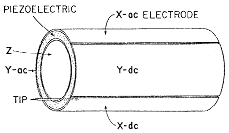
Tube scanner.
Considerable simplification of design and improvement of operating
characteristic were reached by usage of tube scanner, (G. Binnig, D.P.E. Smith.
Rev. Sci. Instrum. 57 (8), 1986, 1688-1689.Full text)which remainds main type scanner up to
now.
Fig 1. Drowing of a tube scanner showing the outside electrode sectuoned
into four equal areas parallel to the axis of the tube. As voltage is applied to
a single outside electrode the tube bends away from that electrode. Voltage
applied to the inside electrode causes a uniform elongation. A small ac signal
and a large dc offset can be sepatated on electrodes 180o apart. A
probe tip is shown mounted for an STM.
Prolytic graphite.
|
|
Highly oriented pyrolytic graphite was introduced in research practice by
Sang-II Park and C.F. Quate (Appl. Phys. Lett. 48 (2), 1986, 112-114. Full text). Its role in scanning probe
microscopy development may not be overestimated. Possibility of comparatively
simply in ambient air to reach atomic resolution have make graphite prevailing
substrate for tunneling microscopy. Area of graphite applications is very wide:
with help of graphite first steps in scanning tunneling microscopy methods
studing are maked, structure for calibrating, atomic flate substrates for
deposition various structures under consideration, good model substance for
theoretical investigations etc.
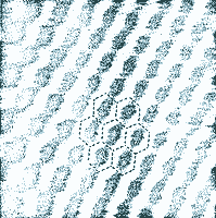
Below one of earlier STM images of graphite
is presented, scan size 20x20 A. |
|
|
Atomic -force microscope.
The Scanning Tunneling Microscope has considerable
restriction, specimens must be electrically conducted. With invention of
scanning atomic-force microscope by Binnig, Quate and
Gerber(Phys.Rev.Lett.56,1986,930-933.Full text.), practicaly all types of specimens (up to
investigation of protein structure in liquids) became available to measuring.
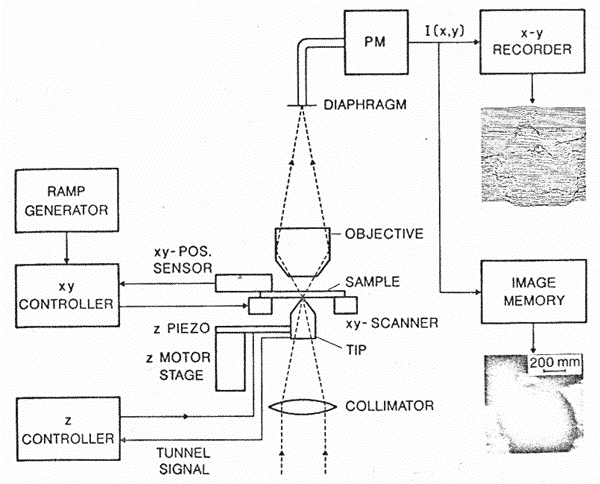
Near-field optical microscope.
Scanning probe microscopy made a major contribution to near-field
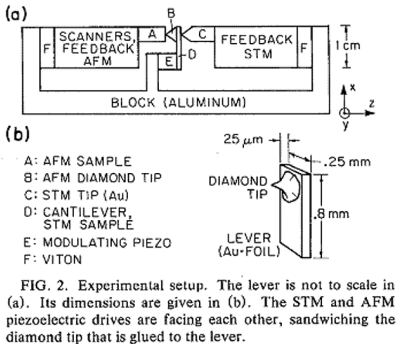
optical microscopy development. Below the block diagram of near-field
optical microscope is presented where to maintaining the probe-surface
gap the tunnel current is used.( Durig U., D. W. Pohl, F. Rohner.Near-field
optical-scanning microscopy.J. Appl. Phys.59 (10).1986. 3318-3327.
Full text.).
|
